Shalby Hospitals, Ahmedabad, India.
Fellowship (South Korea)
Knee
The knee is one of the largest and most complex joints in the body. The knee joins the thigh bone (femur) to the shin bone (tibia). The smaller bone that runs alongside the tibia (fibula) and the kneecap (patella) are the other bones that make the knee joint.
Tendons connect the knee bones to the leg muscles that move the knee joint. Ligaments join the knee bones and provide stability to the knee:

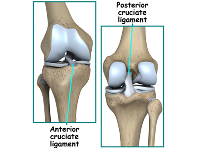
- The anterior cruciate ligament prevents the femur from sliding backward on the tibia (or the tibia sliding forward on the femur).
- The posterior cruciate ligament prevents the femur from sliding forward on the tibia (or the tibia from sliding backward on the femur).
- The medial and lateral collateral ligaments prevent the femur from sliding side to side.
Two C-shaped pieces of cartilage called the medial and lateral menisci act as shock absorbers between the femur and tibia.
Numerous bursae, or fluid-filled sacs, help the knee move smoothly.
COMMON KNEE PROBLEMS
- Chondromalacia patella (also called patellofemoral syndrome): Irritation of the cartilage on the underside of the kneecap (patella), causing knee pain. This is a common cause of knee pain in young people.
- Knee osteoarthritis: Osteoarthritis is the most common form of arthritis, and often affects the knees. Caused by aging and wear and tear of cartilage, osteoarthritis symptoms may include knee pain, stiffness, and swelling.
- Knee effusion: Fluid buildup inside the knee, usually from inflammation. Any form of arthritis or injury may cause a knee effusion.
- Meniscal tear: Damage to a meniscus, the cartilage that cushions the knee, often occurs with twisting the knee. Large tears may cause the knee to lock.
- ACL (anterior cruciate ligament) strain or tear: The ACL is responsible for a large part of the knee's stability. An ACL tear often leads to the knee "giving out," and may require surgical repair.
- PCL (posterior cruciate ligament) strain or tear: PCL tears can cause pain, swelling, and knee instability. These injuries are less common than ACL tears, and physical therapy (rather than surgery) is usually the best option.
- MCL (medial collateral ligament) strain or tear: This injury may cause pain and possible instability to the inner side of the knee.
- Patellar subluxation: The kneecap slides abnormally or dislocates along the thigh bone during activity. Knee pain around the kneecap results.
- Patellar tendonitis: Inflammation of the tendon connecting the kneecap (patella) to the shin bone. This occurs mostly in athletes from repeated jumping.
- Knee bursitis: Pain, swelling, and warmth in any of the bursae of the knee. Bursitis often occurs from overuse or injury.
- Baker's cyst: Collection of fluid in the back of the knee. Baker's cysts usually develop from a persistent effusion as in conditions such as arthritis.
ANTERIOR CRUCIATE LIGAMENT (ACL)
Athletes place the the anterior cruciate ligament of the knee under a tremendous amount of stress. If left untreated, ACL injuries can lead to cartilage damage. ACL injuries are far more common in young women athletes than in men, but we don't know why this is so.
ACL surgery involves the reconstruction of the ligament. While numerous ACL surgery techniques exist, Dr. Dhaval Sagala will recommend the best approach based on your injuries and condition – to return you the full range of motion and stability to the knee. Dr Dhaval Sagala has treated Large Number of ACL injuries Cases.
Post-surgery, our rehabilitation specialists and athletic trainers will take you through a comprehensive rehabilitation plan for your quick and healthy recovery.
Do you have an ACL injury?
ACL Causes of Injury
The ACL is the most commonly injured knee ligament. It tears during sudden dislocation, torsion, or hyperextension of the knee. Swelling attributed to bleeding from the tear may cause pressure and some discomfort, but the torn ligament itself rarely causes much pain.
Due to the lack of severe pain most patients are able to postpone surgery based on the extent of the damage to other tissues in the knee. It is a very common injury in football, hockey, skiing, skating, soccer (and other field sports) and in most cases basketball, due to the enormous amount of pressure, weight, and torque the knee must withstand.
Usually the injury occurs when someone tries to rapidly change direction with the leading leg out, twisting the knee; this is usually followed by a loud popping sound. The injury may also occur under sudden high pressure contact, especially side on. Generally if the knee is locked, and the leg is firmly planted, then there is a much greater risk of injury.
ACL ruptures can be caused by these factors:
Environmental: Sports which include running, jumping, and landing pose the most potential for injury to the athlete. Interestingly, the risk for rupture of the anterior cruciate ligament does not increase in contact sports (as opposed to noncontact sports). About 70 percent of ACL tears are noncontact injuries.
Anatomical: ACL injuries are especially common in female athletes, due to many possible contributing factors. The most prevalent explanation proposes that female athletes tend to land more straight-legged than men, removing the quadriceps' muscles shock-absorbing action on the knee. Often the knee on a straight leg can't withstand this and bends sideways.
Hormonal: High levels of hormones including estrogen have been associated with an increased risk of ACL rupture.
Signs Include:
- Giving way of the knee
- A popping sound at the time of injury
- Swelling of the knee joint
- Pain in the knee
- Surgen will examine you, conduct physical tests and take X-rays to determine the extent of damage to your ACL.
- Treatment for an ACL injury
- Nonsurgical Options
Dr. Dhaval Sagala recommending the nonsurgical option may specify physical therapy, the wearing of a knee brace, or adapting some typical activities. Physical therapy may be recommended to strengthen muscles around the knee – in order to to compensate for the absence of a healthy ACL. Physical therapy will focus on strengthening muscles such as the hamstring, quadriceps, calf, hip, and ankle. This therapy will help to re-establish a full range of motion of the knee.
People applying nonsurgical options can expect to be back to normal daily activity within one month.
Other non-surgical options include prolotherapy, which has been shown by Reeves in a small RCT to reduce translation on KT-1000 arthrometer versus placebo
Surgical Options Dr. Dhaval Sagala may recommend reconstructive surgery if the knee gives way during typical daily activities, shows functional instability, or if the person continues to enjoy high-risk activities.
Reconstructive surgery may also be recommended if there is damage to the meniscus (cartilage). This surgery is completed using arthroscopic techniques. There is also an option for an autograft to be done using a chosen tendon.
You will want to weigh the pros and cons of surgical treatment. Consider possible complications and talk with your surgeon before proceeding with this form of treatment.
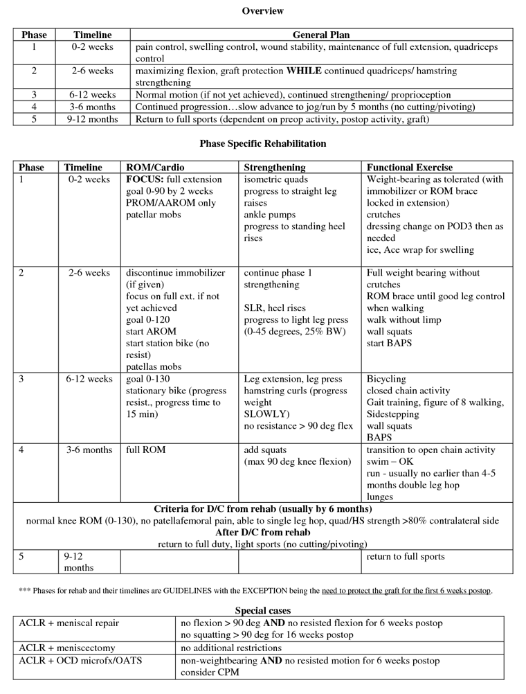
If surgery is your chosen path, there are rehabilitation requirements to consider. Physical therapy must be completed in three phases after the surgery is completed. With the use of the surgical treatment option, rehabilitation included, a patient can expect to be returning to previous and desired levels of activity in six to nine months
ACL Surgical Technique
Surgen replaces the damaged ACL with strong, healthy tissue taken from another area near your knee. You might utilize a strip of tendon from under your kneecap (patellar tendon) or your hamstring. Surgeon threads the tissue through the inside of your knee joint and secures the ends to your thighbone and shinbone. In a few cases when the ACL is torn cleanly from the bone, it can be repaired. Less active people may be treated non-invasively with a program of muscle strengthening rather than knee surgery.
ACL Post OP Rehabilitation Program